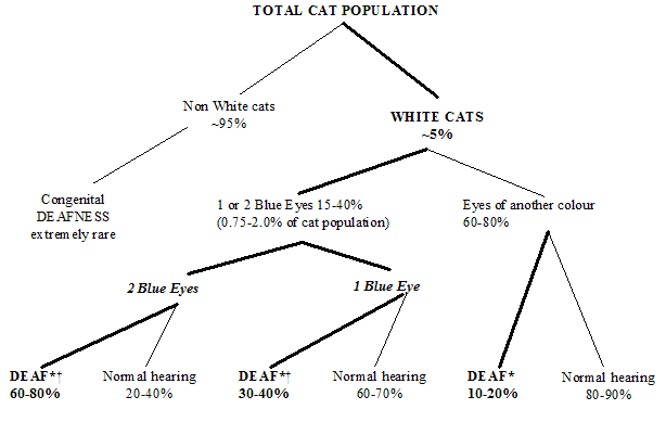
*DEAFNESS may be unilateral or bilateral.
† These two groups of cats account for 0.25-1.5% of the cat population.
From this it may be determined that if a cat has 2 blue eyes, it is 3-5 times more likely to be deaf than a cat with 2 coloured eyes (non-blue), and a cat with 1 blue eye is twice as likely to be deaf than the cat with 2 coloured eyes (non-blue). In addition, while longhaired and short-haired cats are just as likely to be unilaterally deaf, longhaired cats are 3 times as likely to be bilaterally deaf.
In a feral situation deaf white cats experience strong negative natural selection pressure as, in addition to being deaf, photophobic and having reduced vision in low light conditions, they are also believed to have reduced resistance to disease and semi-sterility. However, studies of pet cats have shown that there are far more white cats than would be expected and the reason for the high prevalence appears to be human preference and intervention. Many cat breeds are known to have the W gene, and can, therefore, produce deaf white individuals. A number of breeds now insist of white cats being checked for deafness (e.g. using BAER testing - auditory evoked potentials), and do not allow deaf white cats to be bred from, e.g. Norwegian Forrest Cat.
Delack J B (1984) Hereditary deafness in the white cat. Compendium on continuing Education for the practising Veterinarian 6, 609 – 617 (abstract above)
Heid S, Hartmann R, Klinke R. (1998) A model for prelingual deafness, the congenitally deaf white cat - population statistics and degenerative changes, Hearing Research 115:101-112
Bergsma, D.R & Brown, K.S (1971) White fur, blue eyes and deafness in the domestic cat, Journal of Heredity 62:171-18
Strain GM. Deafness in blue-eyed white cats: the uphill road to solving polygenic disorders. Vet J. 2007;173(3):471-2
Ataxia – uncoordinated walking
There are a number of papers that report ataxia (uncoordinated walking) in young domestic shorthaired cats. However, while affected cats may present with similar clinical signs they probably represent a number of different conditions with differing modes of inheritance (most of which are unknown). For example: cats have been seen with cerebellar abiotrophy (which results from spontaneous premature death of brain cells) where affected cats developed severe ataxia, wide-based stance, symmetrical hypermetria (high stepping walk) and spasticity (poorly controlled movements) especially affecting forelimbs. Whole body tremors, intention tremor of the head and postural reaction deficits were also present. Ophthalmic exam revealed vertical nystagmus (rapid sideways movement of the eyes), bilateral absent menace responses, decreased pupillary light reflexes and signs of end-stage retinal degeneration (diffuse tapetal hyperreflectivity and depigmentation of the nontapetal fundus). Clinical signs were slowly progressive with onset of gait disorder and visual deficits (blindness) at 1.5 years and 3 years of age respectively. Pathology revealed cerebellar cortical abiotrophy (cerebellum 2/3rds the normal size) with marked reduction in number of Purkinje cells and attenuation of the granule cell layer. The Purkinje cells were replaced by astrocytes. The ocular changes included selective photoreceptor degeneration with marked reduction in the number of retinal rod and cone cells (Barone et al 2002). Other cases have presented at an earlier age (7-9 weeks), but developed similar clinical signs, including a wide-based stance, ataxia, hypermetria and intention tremor. In that case the pathological lesions consisted of multifocal Purkinje cell degeneration with reactive gliosis and cerebellar foliar disorganization, and lesions were also seen in the medulla and spinal cord. The condition was suspected to have a recessive mode of inheritance as it related to sibling interbreeding (Willoughby & Kelly 2002). Cerebellar degeneration has been seen in a number of cats spanning 2 generations. Magnetic resonance imaging indicated that size of the cerebellum of diseased cats was markedly reduced. Pathology showed cerebellar cortical degeneration, with extensive destruction of Purkinje cells. The condition was shown to have autosomal recessive mode of inheritance (Inada et al 1996). Cerebellar hypoplasia (which results from a lack of growth of the cerebellum) has been seen in 3 litter mates, with signs of dysmetria (difficulty in walking), intention tremors, muscular hypertonia (increased muscle tone) and multiple abnormalilities in postural reflexes being seen shortly after birth.
Neuroaxonal Dystrophy is a degenerative neurological disorder characterized by swelling of the distal segments of axons within the central nervous system. It is similar to the syndrome of feline hereditary neuroaxonal dystrophy (FHND). However, in 1 case series affected cats had no inner ear involvement and were affected at a later age (6-9 months) compared to FHND, and abnormal coat colour or dilution was not a consistent feature. Clinical signs consisted of sudden onset hindlimb ataxia progressing to hindlimb paresis and paralysis. Affected cats were DSH, all siblings with the same queen from several litters. Pathologically, there was marked ballooning of axonal processes, with spheroid formation and vacuolation in specific regions of the brain and spinal cord (Carmichael et al 1993). The syndrome in this report differs from the previously described FHND in that no inner ear involvement was seen and onset of clinical signs occurred at a later age. In addition, although some of the affected cats did have diluted coat colors, abnormal coat color was not always associated with clinical disease. Feline hereditary neuroaxonal dystrophy (FHND) is characterized clinically by an abnormal coat colour and development of progressive ataxia during infancy. Breeding experiments indicate that the disease is inherited in an autosomal recessive manner. The most prominent microscopic alterations were marked ballooning of nerve cell processes within specific regions of the brain stem and atrophy of the cerebellar vermis. Examination of the inner ears revealed depletion of neurons in the spiral ganglia and homogeneous eosinophilic bodies within the spiral ganglia, nerve fibre tracts and organ of Corti.
Barone, G., Foureman, P., deLahunta, A. (2002): Adult-onset cerebellar cortical abiotrophy and retinal degeneration in a domestic shorthair cat, Journal of the American Animal Hospital Association 38:51-54
Carmichael, K.P et al (1993) Neuroaxonal Dystrophy in a Group of Related Cats, Journal of Veterinary Diagnostic Investigation 5:585-590
Inada S, Mochizuki M, Izumo S, Kuriyama M, Sakamoto H, Kawasaki Y,Osame M. (1996) Study of hereditary cerebellar degeneration in cats. Am J Vet Res. 57(3):296-301
Scheidy SF (1953) Familial cerebellar hypoplasia in cats, North American Veterinarian 34:118-119
Woodard JC, Collins GH, Hessler JR. (1974) Feline hereditary neuroaxonal dystrophy. Am J Pathol. ;74(3):551-66
Willoughby, K., Kelly, D.F. (2002): Hereditary cerebellar degeneration in three full sibling kittens, Veterinary Record 151:295-298
Lysosomal storage diseases – also see other sections
Lysosomes are structures found within cells that contain enzymes involved in metabolism of cellular products. Lysosomal storage disorders arise when the lysosomes are deficient in an essential enzyme, which leads to a build-up (storage) of a product within cells. An autosomal recessive mode of inheritance has been identified for many of these diseases. The diseases are extremely rare, which may result in them being under-recognised. In addition, confirmation of the enzyme deficiency is often extremely difficult, especially ante-mortem (before death), and specialist laboratory techniques are usually required. Certain disorders have been associated with specific breeds, but all the disorders have also been identified in domestic shorthair and/or longhaired cats (Mucopolysaccharidosis VII, Niemann-Pick Type C, globoid leukodystrophy and mucolipidosis). The majority of conditions show clinical signs from an early age, and neurological abnormalities are a frequent finding. Enlargement of the liver, stunted growth and ocular abnormalities are also frequently identified. Genetic tests are available for some conditions, and detection of abnormal metabolites in the urine may help to support the diagnosis of some conditions. more |